43 respiratory system diagram without labels
30 Fun And Interesting Respiratory System Facts For Kids The Respiratory Tract Image: Shutterstock The respiratory tract is divided into two sections, namely, upper and lower. The part above the voice box or larynx is upper respiratory tract and the one below it is lower respiratory tract. The respiratory tract is lined by respiratory mucosa or respiratory epithelium (2). Lymphatic System: Lymphatic Functions, Diagram at Embibe The lymphatic ducts are of the following two kinds: 1. Thoracic Duct The lymphatic vessels of the left side unite to form a thoracic duct which begins at the cisterna chyli (a sac-dilation situated in front of the first and second number vertebrae). The thoracic duct contains several valves that discharge its lymph into the left subclavian vein.
Bronchioles: Anatomy, Function, and Treatment - Verywell Health The function of the bronchioles is to deliver air to a diffuse network of around 300 million alveoli in the lungs. 5 As you inhale, oxygenated air is pulled into the bronchioles. Carbon dioxide collected by the alveoli is then expelled from the lungs as you exhale. The bronchioles are not inert. The smooth muscles that surround the airways ...
Respiratory system diagram without labels
Bronchial Branches - anatomical labeling computational anatomy based on ... Bronchial Branches - 100 images - nomenclature of branches of the bronchial artery in pig download, anatomy of the bronchi anatomy drawing diagram, download stl file bronchial tree 3d printing model design to 3d print, locate portions of aorta, How Lungs Work - American Lung Association The WINDPIPE (trachea) is the passage leading from your throat to your lungs. The windpipe divides into the two main BRONCHIAL TUBES, one for each lung, which divides again into each lobe of your lungs. These, in turn, split further into bronchioles. Lungs and Blood Vessels Your right lung is divided into three LOBES, or sections. Anatomical Line Drawings - Medscape SELECT LINE DRAWING. Arterial Supply - anterior view. go to drawing with labels go to drawing without labels. Digestive System - anterior view. go to drawing with labels go to drawing without ...
Respiratory system diagram without labels. Upper Respiratory Tract System - upper respiratory tract quiz ... Upper Respiratory Tract System - 17 images - human respiratory system and alveolus structure model human body, m altman presentation ch 7 respiratory system drugs, respiratory system online presentation, diseases and conditions of the respiratory system basicmedical key, Mechanics of Breathing - Inspiration - TeachMePhysiology Inspiration is the phase of ventilation in which air enters the lungs. It is initiated by contraction of the inspiratory muscles: Diaphragm - flattens, extending the superior/inferior dimension of the thoracic cavity. External intercostal muscles - elevates the ribs and sternum, extending the anterior/posterior dimension of the thoracic cavity. Circulatory System Diagram - New Health Advisor There are different types of circulatory system diagrams; some have labels while others don't. The color blue stands for deoxygenated blood while red stands for blood which is oxygenated. Below you'll see diagram specified to the heart, as well as circulatory system diagram of the whole body: How Does the Human Circulatory System Work? 1. Heart WHMIS 2015 - Pictograms : OSH Answers Pictograms are graphic images that immediately show the user of a hazardous product what type of hazard is present. With a quick glance, you can see, for example, that the product is flammable, or if it might be a health hazard. Most pictograms have a distinctive red "square set on one of its points" border.
Bronchi: Anatomy, function and histology - Kenhub Need a refresher on the basic anatomy of the respiratory system before diving into all things bronchi related? Revise with our respiratory system quizzes and labeled diagrams. Bronchopulmonary segment. ... which is an advantage during surgery since a bronchopulmonary segment can be removed without affects other nearby segments. There are 10 ... Respiratory And Digestive System Diagram - o l sinhala science lesson ... Respiratory And Digestive System Diagram - 16 images - structures of the mediastinum lateral view 200049 02a, body systems the internets best learning resource diagram diagram of, anatomy lab test 2 flashcards quizlet, the respiratory and digestive systems stock image c020 1161, Upper Respiratory Tract Anatomy - upper respiratory tract anatomy ... Upper Respiratory Tract Anatomy - 15 images - anat2241 respiratory system embryology, respiratory system anatomy larynx to lung model youtube, the respiratory system anatomy and physiology questions proprofs quiz, sore throat with throat swollen closeup open mouth with posterior, EOF
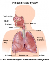
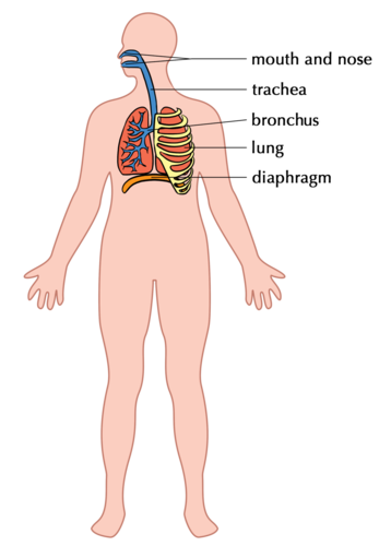
Sternum: Anatomy, parts, pain and diagram | Kenhub The sternum is the bone that lies in the anterior midline of our thorax. It forms part of the rib cage and the anterior-most part of the thorax. Its functions are to protect the thoracic organs from trauma and also form the bony attachment for various muscles. It is also the center around which the superior 10 ribs directly or indirectly ... Important Question for Class 10 Science Life Processes - Learn CBSE Respiratory pigment present in human beings is haemoglobin. Rings of cartilage are C in shape, stacked one on top of the other. These cartilaginous rings prevent the trachea from collapsing and blocking the airway. Question 23. (a) Draw a diagram of human respiratory system and label: Trachea, Bronchi and Diaphragm. (b) Give reasons for the ... Anatomical Line Drawings - Medscape SELECT LINE DRAWING. Arterial Supply - anterior view. go to drawing with labels go to drawing without labels. Digestive System - anterior view. go to drawing with labels go to drawing without ... How Lungs Work - American Lung Association The WINDPIPE (trachea) is the passage leading from your throat to your lungs. The windpipe divides into the two main BRONCHIAL TUBES, one for each lung, which divides again into each lobe of your lungs. These, in turn, split further into bronchioles. Lungs and Blood Vessels Your right lung is divided into three LOBES, or sections.
32 Label The Respiratory System Quiz - Labels 2021
Bronchial Branches - anatomical labeling computational anatomy based on ... Bronchial Branches - 100 images - nomenclature of branches of the bronchial artery in pig download, anatomy of the bronchi anatomy drawing diagram, download stl file bronchial tree 3d printing model design to 3d print, locate portions of aorta,
Respiratory system - Teaching resources
Neuron B&w Clip Art at Clker.com - vector clip art online, royalty free & public domain

Respiratory System printable diagram pdf – Science A Plus
Label Respiratory System Diagram - ClipArt Best
Respiratory system Archives - Medical Information Illustrated

Pinterest • The world’s catalog of ideas

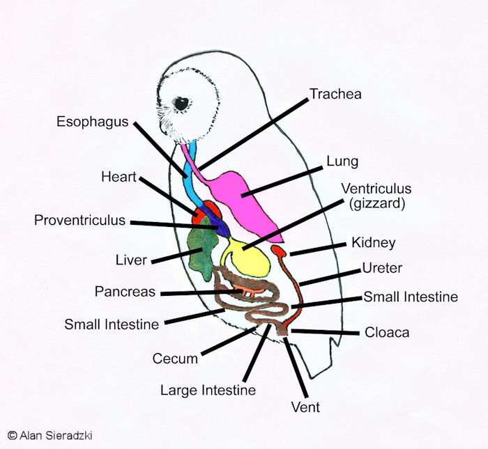
Fetal Pig Anatomy Quiz

Respiratory system diagram to label | Teaching Resources
Respiratory System Diagram To Label - Pensandpieces

Blank Ear Diagram | Human ear diagram, Ear anatomy, Ear diagram
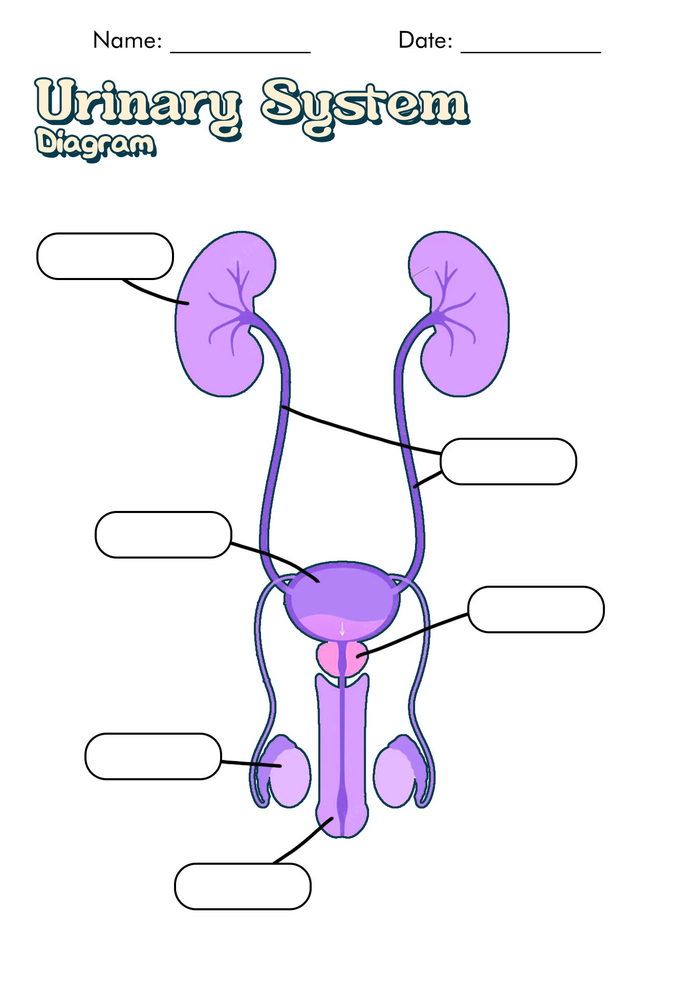
14 Best Images of Urinary System Worksheets - Blank Urinary System, Urinary System Anatomy and ...

Respiratory System With Label Drawing at GetDrawings.com | Free for personal use Respiratory ...

Easy Respiratory System Diagram Class 10 - Diagram Media

Circulatory System By: Connor

Unlabelled Respiratory System Clip Art at Clker.com - vector clip art online, royalty free ...
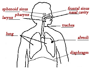
Diagram Of Human Respiratory System With Labels

Respiratory System Clip Art at Clker.com - vector clip art online, royalty free & public domain
Post a Comment for "43 respiratory system diagram without labels"