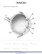
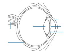
44 picture of the eye with labels
Label Eye Printout - EnchantedLearning.com Label the Eye Diagram. Human Anatomy. Read the definitions, then label the eye anatomy diagram below. Cornea - the clear, dome-shaped tissue covering the front of the eye. Iris - the colored part of the eye - it controls the amount of light that enters the eye by changing the size of the pupil. Lens - a crystalline structure located just behind ... Label Parts of the Human Eye - University of Dayton Parts of the Eye. Select the correct label for each part of the eye. The image is taken from above the left eye. Click on the Score button to see how you did. Incorrect answers will be marked in red. ...
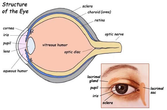
Eye Anatomy Detail Picture Image on MedicineNet.com Picture of Eye Anatomy Detail The eye is our organ of sight. The eye has a number of components which include but are not limited to the cornea, iris, pupil, lens, retina, macula, optic nerve, choroid and vitreous. Cornea: clear front window of the eye that transmits and focuses light into the eye.

Picture of the eye with labels
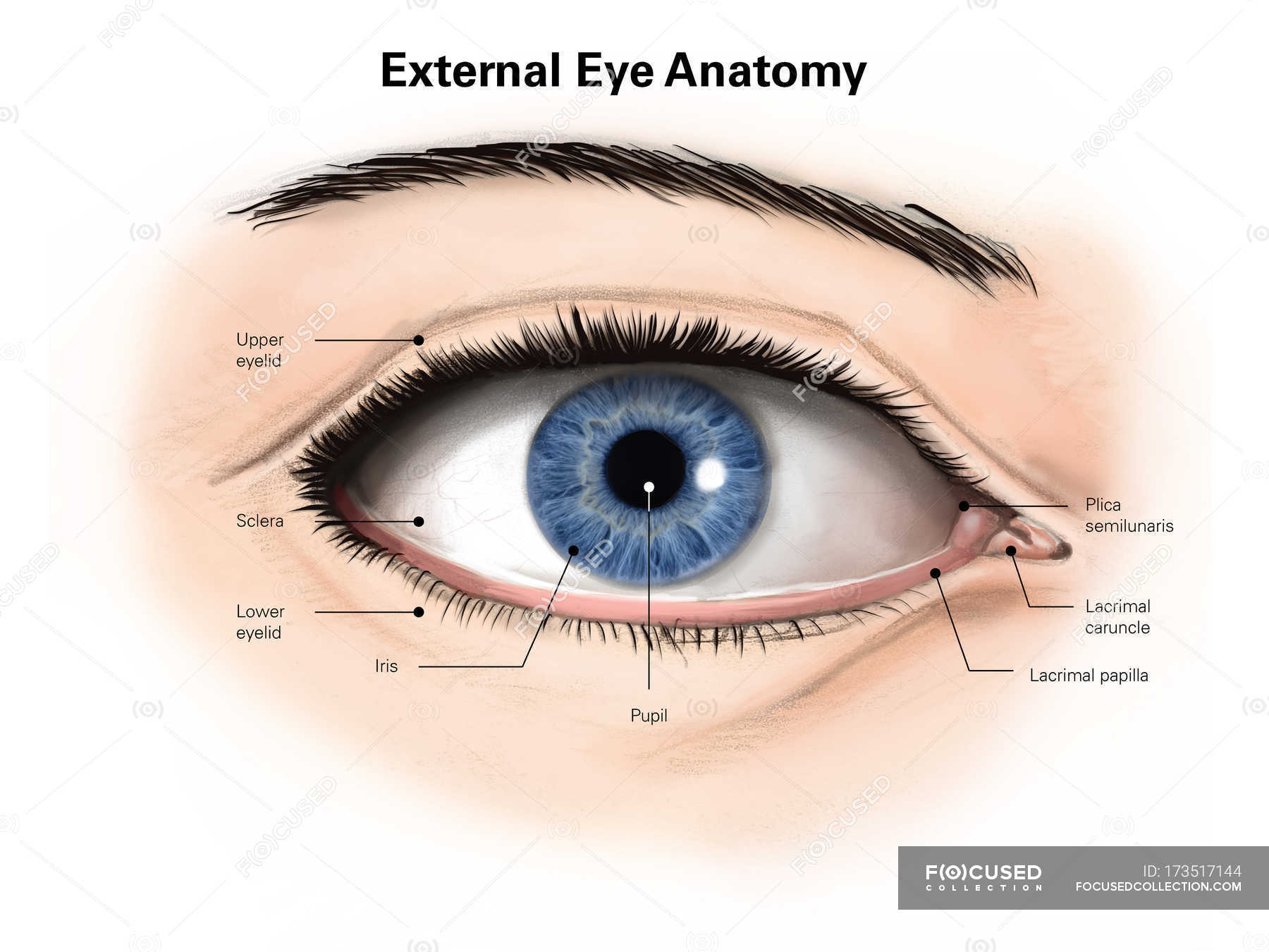
Labelling the eye — Science Learning Hub In this interactive, you can label parts of the human eye. Use your mouse or finger to hover over a box to highlight the part to be named. Drag and drop the text labels onto the boxes next to the eye diagram If you want to redo an answer, click on the box and the answer will go back to the top so you can move it to another box. Eye Anatomy: Parts of the Eye and How We See Behind the anterior chamber is the eye's iris (the colored part of the eye) and the dark hole in the middle called the pupil. Muscles in the iris dilate (widen) or constrict (narrow) the pupil to control the amount of light reaching the back of the eye. Directly behind the pupil sits the lens. The lens focuses light toward the back of the eye. 31 Most Beautiful Eyes in the World - Woman's World 31 People With the Most Striking Eyes in the World. By Jaclyn Anglis August 31, 2021. We can't help but see beautiful eyes and smile, can you? Beautiful eyes come in so many different colors, shapes, and sizes. But no matter what gorgeous form they take, all stunning eyes have one thing in common: They're guaranteed to make people stop in ...
Picture of the eye with labels. PDF Eye Anatomy Handout - National Institutes of Health of light entering the eye. Lens: The lens is a clear part of the eye behind the iris that helps to focus light, or an image, on the retina. Macula: The macula is the small, sensitive area of the retina that gives central vision. It is located in the center of the retina. Optic nerve: The optic nerve is the largest sensory nerve of the eye. Label the Eye - The Biology Corner Label the Eye Shannan Muskopf December 30, 2019 This worksheet shows an image of the eye with structures numbered. Students practice labeling the eye or teachers can print this to use as an assessment. There are two versions on the google doc and pdf file, one where the word bank is included and another with no word bank for differentiation. What is an eye mark and why do I need it? - Consolidated Label An 'eye mark' (also known as 'eye spot') is a small rectangular printed area located near the edge of the printed flexible packaging material. A sensor on the form-fill-seal (FFS) machine reads the eye mark to identify packaging material, control the material's position, and coordinate the separation and cutting of the flexible packaging material. Transverse section of eye anatomy with labels. - Getty Images View top-quality illustrations of Transverse Section Of Eye Anatomy With Labels. Find premium, high-resolution illustrative art at Getty Images.
Labelled Diagram of Human Eye, Explanation and Function - VEDANTU The human eye is a part of the sensory nervous system. Labeled Diagram of Human Eye The eyes of all mammals consist of a non-image-forming photosensitive ganglion within the retina which receives light, adjusts the dimensions of the pupil, regulates the availability of melatonin hormones, and also entertains the body clock. Eye Pictures, Anatomy & Diagram | Body Maps - Healthline Eyes are approximately one inch in diameter. Pads of fat and the surrounding bones of the skull protect them. The eye has several major components: the cornea, pupil, lens, iris, retina, and sclera. Eye Anatomy: A Closer Look At the Parts of the Eye - All About Vision The eye's crystalline lens is located directly behind the pupil and further focuses light. Through a process called accommodation, this lens helps the eye automatically focus on near and approaching objects, like an autofocus camera lens. ... The retina acts like an electronic image sensor of a digital camera, converting optical images into ... 10 Amazing Label Eye Parts and The Function - label template Label eye parts: The retina The retina restores light into electrical stimulations that are then transferred to your brain via nerve fibers and processed into images. The lens To let you perceive objects, the lens concentrates incoming light onto a single point on your retina. The Pupil The spot in your eye where light can enter.
Label Functions of Parts of the Human Eye - University of Dayton Functions of the Parts of the Eye. Select the correct label for the function of each part of the eye. The image is taken from above the left eye. Click on the Score button to see how you did. Incorrect answers will be marked in red. Label the Eye Worksheet - Teacher-Made Learning Resources - Twinkl In this resource, you'll find a 2-page PDF that is easy to download, print out, and use immediately with your class. The first page is a labelling exercise with two diagrams of the human eye. One is a view from the outside, and the other is a more detailed cross-section. Challenge learners to label the parts of the eye diagram. On the second page, you'll find a set of answers showing ... Eye Anatomy: 16 Parts of the Eye & Their Functions - Vision Center The lens of the eye (or crystalline lens) is the transparent lentil-shaped structure inside your eye. This is the natural lens. It is located behind the iris and to the front of the vitreous humor (vitreous body). The vitreous humor is a clear, colorless, gelatinous mass that fills the gap between the lens and the retina in the eye. Quiz: Label The Parts Of The Eye - ProProfs Quiz Quiz: Label The Parts Of The Eye. Do you know the anatomy of the human eye very well? Can you label the parts of the eye in the quiz below? Give it a try and evaluate yourself. The eye has many important parts, each with different functions, including the cornea, pupil, sclera, and many more. Can you tell where these parts are located and what ...
My Life in Tupperware Bins: The Life of a Tall Girl
Human Eye Diagram - Human Body Pictures & Images - Science for Kids Photo description: This human eye diagram gives an excellent overview of the human eye. The cross section features labeled parts such as the iris, pupil, cornea, lens, retina, choroid, optic disc, optic nerve and fovea. For more information on eyes, check out our range of interesting human eye facts.

Amazing close-up photos of animal eyes (9 pics) | Amazing Creatures
Human Eye Anatomy Pictures, Images and Stock Photos Vector illustration of the structure of the eye. Anatomy of the... Structure of the eye, parts of the eye. Retina, macula, blind spot, optic nerve, cones, rods, vitreous humor, ciliary body, lens, pupil, aqueous humor, cornea, iris, sclera, choroid. Eye layers The inner layer of the eye, or retina, is similar to film in a camera.

World's Beautiful things around us !: Beautiful nature| Eye cooling pictures every body loves to ...
A Picture of the Eye - WebMD The front part (what you see in the mirror) includes: Iris: the colored part. Cornea: a clear dome over the iris. Pupil: the black circular opening in the iris that lets light in. Sclera: the ...

Kate McKinnon Hot Pics and Bio | Picture Perfect
The Human Eye (Eyeball) Diagram, Parts and Pictures The eyeball is a round gelatinous organ that contains the actual optical apparatus. It is approximately 25 mm in diameter and sits snugly in the orbit where six muscles control its movement. The eyeball has three layers, each of which has several important structures that are essential for the sense of vision. Wall of the Eyeball
32 Label The Eye Quiz - Labels 2021
Human Eye Coloring Page | crayola.com The eye is the organ that collects images and sends them to the brain, so you can see. The eye is protected by the bones of your skull and six muscles. Light comes through the pupil which causes the cornea and lens to focus on an image. When the image is projected through the eye, onto the retina wall, the image appears upside down. Eye ...
Label the Part of the Eye -- Exploring Nature Educational Resource
Eye Diagram With Labels and detailed description - BYJUS A brief description of the eye along with a well-labelled diagram is given below for reference. Well-Labelled Diagram of Eye The anterior chamber of the eye is the space between the cornea and the iris and is filled with a lubricating fluid, aqueous humour. The vascular layer of the eye, known as the choroid contains the connective tissue.
Human eye with labels — retina, human organ - Stock Photo | #173517144
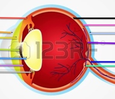
Solved B с A E F D Match the following parts of the eye with - Chegg Anatomy and Physiology questions and answers. B с A E F D Match the following parts of the eye with the labels in the picture above. A Iris F Cornea В. Ciliary Muscles G Optic Nerve C Lens E Retina Aqueous and Vitreous Fluid. Question: B с A E F D Match the following parts of the eye with the labels in the picture above.

The BioLogs: CSEC - The Eye - functions of the various parts
Eye Anatomy Diagram - EnchantedLearning.com Definitions : Aqueous humor - the clear, watery fluid inside the eye. It provides nutrients to the eye. Astigmatism - a condition in which the lens is warped, causing images not to focus properly on the retina. Binocular vision - the coordinated use of two eyes which gives the ability to see the world in three dimensions - 3D.

32 Label Eye Parts - Labels Design Ideas 2020
PDF Parts of the Eye - National Institutes of Health Eye Diagram Handout Author: National Eye Health Education Program of the National Eye Institute, National Institutes of Health Subject: Handout illustrating parts of the eye Keywords: parts of the eye, eye diagram, vitreous gel, iris, cornea, pupil, lens, optic nerve, macula, retina Created Date: 12/16/2011 12:39:09 PM

Quiz – Page 2
Diagram of the Eye - Lions Eye Institute Instructions Click the parts of the eye to see a description for each. Hover the diagram to zoom. Need any help? If you would like to know more about us, or want to make an appointment, please don't hesitate to get in touch. (08) 9381 0777 carecentre@lei.org.au Request an appointment Customer Care Centre (08) 9381 0777

LabelRIGHT Ultimate for Windows Bar Code Label Design and Printing Software - Worth Data
1,109,890 Human eye Images, Stock Photos & Vectors - Shutterstock Human eye royalty-free images 1,109,890 human eye stock photos, vectors, and illustrations are available royalty-free. See human eye stock video clips Image type Orientation People Artists Sort by Popular Biology Healthcare and Medical Icons and Graphics human eye macro photography anatomy iris eye 3d rendering pupil Next of 11,099

Fitness and Bodybuilder Model Frank DeFeo | Model Galleries
31 Most Beautiful Eyes in the World - Woman's World 31 People With the Most Striking Eyes in the World. By Jaclyn Anglis August 31, 2021. We can't help but see beautiful eyes and smile, can you? Beautiful eyes come in so many different colors, shapes, and sizes. But no matter what gorgeous form they take, all stunning eyes have one thing in common: They're guaranteed to make people stop in ...

KATMAN SCIENCE
Eye Anatomy: Parts of the Eye and How We See Behind the anterior chamber is the eye's iris (the colored part of the eye) and the dark hole in the middle called the pupil. Muscles in the iris dilate (widen) or constrict (narrow) the pupil to control the amount of light reaching the back of the eye. Directly behind the pupil sits the lens. The lens focuses light toward the back of the eye.
31 Label The Eye Quiz - Best Labeling Ideas
Labelling the eye — Science Learning Hub In this interactive, you can label parts of the human eye. Use your mouse or finger to hover over a box to highlight the part to be named. Drag and drop the text labels onto the boxes next to the eye diagram If you want to redo an answer, click on the box and the answer will go back to the top so you can move it to another box.
+[%232]+(2).jpg)
Aquarious' TLC Fan Blog: Lisa Lopes & Hype Williams' 'Nature Girl' Photo Shoot
picture front of the eye without labels clipart 20 free Cliparts | Download images on Clipground ...

Fitness and Bodybuilder Model Frank DeFeo | Model Galleries
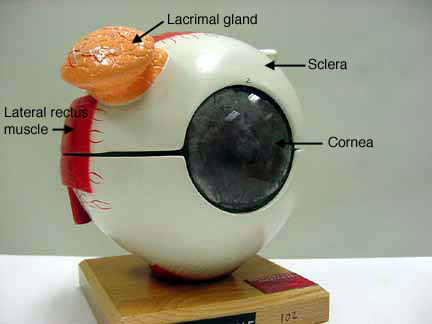
Apparently not even the professor knows what this part of the eye is. The tan lump in this model ...
Post a Comment for "44 picture of the eye with labels"